Large imaging study changes understanding of the origins of Parkinson’s rest tremor
| A Finnish clinical imaging study shows that rest tremor in Parkinson’s disease is not explained by greater dopamine loss. In contrast, tremor appears to be associated with relatively better-preserved dopamine function. |
|
|
|
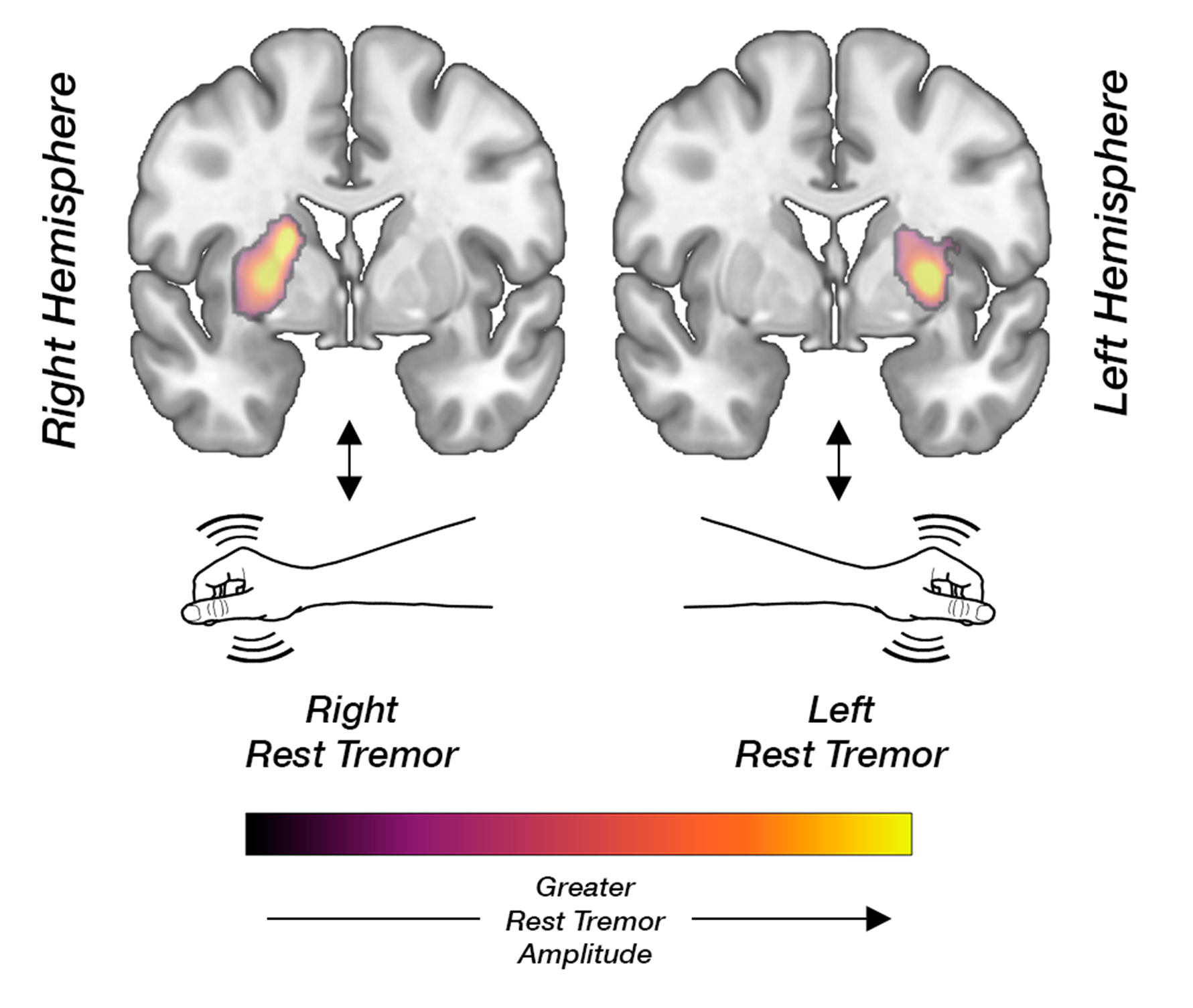
Researchers from the University of Turku and Turku University Hospital, Finland, analysed clinical data and dopamine transporter (DAT) imaging data from 414 Finnish patients. The cohort consisted of patients examined in routine clinical practice for uncertain parkinsonism or tremor, making the findings exceptionally well generalisable to real-world clinical settings. The results were published on 19 March 2026 in Neurology®, the prestigious medical journal of the American Academy of Neurology. The cardinal motor symptoms of Parkinson’s disease are slowness of movement (bradykinesia), muscle stiffness (rigidity), and rest tremor. Bradykinesia and rigidity are known to reflect degeneration of dopamine-producing neurons. Because most brain pathways cross, this association is typically observed in the striatum on the side opposite to the symptoms. In contrast, the biological basis of rest tremor has long remained uncertain. The study revealed a clear and consistent phenomenon: rest tremor was associated with higher dopamine transporter binding in the striatum on the same side as the tremor. Other cardinal motor symptoms, however, showed the expected correlation with dopamine deficits in the opposite hemisphere. “These results show that more severe rest tremor is not simply a marker of more advanced damage to the dopamine system,” says the lead author, Neurologist Kalle Niemi, MD, PhD. “Tremor appears to involve a partly distinct neurobiological mechanism.” The findings confirm the group’s earlier observations made using data from the international Parkinson’s Progression Markers Initiative (PPMI) cohort, where a novel imaging analysis technique developed by the research team was first applied. The replication of the results in an independent and clinically representative cohort strengthens the reliability of the observed phenomenon. “Our findings support the view that different symptoms of Parkinson’s disease may be driven by partly distinct neural network and neurotransmitter mechanisms,” Niemi explains. “This may help explain why tremor behaves differently from symptoms such as bradykinesia.” Using the same methodological framework, the research team also demonstrated that key non-motor symptoms of Parkinson’s disease including depression, anxiety, and REM sleep behaviour disorder are primarily linked to monoaminergic systems other than dopamine. Taken together, these findings reinforce the concept of Parkinson’s disease as a complex brain disorder involving alterations across multiple neural networks and neurotransmitter systems. A more precise understanding of the biological differences between symptoms may, in the future, enable the development of more targeted and personalised treatment approaches. |